
Dosimetric Comparison of Doses to Organs at Risk Using 3-D Conformal Radiotherapy versus Intensity Modulated Radiotherapy in Postoperative Radiotherapy of Periampullary Cancers: Implications for Radiation Dose Escalation
Abstract
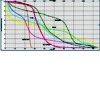
Context Postoperative periampullary cancers with high risk features are managed with adjuvant chemo radiotherapy. Doses of 40-50 Gy have generally been used in conventional radiotherapy. Dose escalation with conventional radiotherapy has been restricted due to surrounding critical organs. Objective The objective of this dosimetric analysis was to evaluate the dose of radiation received by organs at risk using 3D conformal radiotherapy (3DCRT) and intensity modulated radiotherapy (IMRT). Methods Ten postoperative patients of periampullary cancers were selected for this dosimetric analysis. Planning CT scans films were taken with slice thickness of 2.5 mm and transferred to EclipseTM treatment planning system. The clinical target volume (CTV) included the postoperative tumor bed and draining lymph nodal areas. A 1 cm margin was taken around the CTV to generate the planning target volume (PTV). Critical structures contoured for evaluation included bowel bag, bilateral kidneys, liver, stomach and spinal cord. IMRT plans were generated using seven field coplanar beams and 3DCRT planning was done using one anterior and two lateral fields. A dose of 45 Gy in 25 fractions was prescribed to the PTV. Results V45 for bowel bag was 212.3±159.0 cc (mean volume ± standard deviation) versus 80.9±57.4 cc in 3DCRT versus IMRT (P=0.033). The V28 dose analysis for bilateral kidneys showed a value of 32.7±23.5 cc (mean volume ± standard deviation) versus 7.9±7.4 cc for 3DCRT versus IMRT, respectively (P=0.013). The D60 for liver using 3DCRT and IMRT was 28.4±8.6 Gy (mean dose ± standard deviation) and 19.9±3.2 Gy, respectively (P=0.020). Conclusions Doses to bowel bag, liver and kidneys was significantly reduced using IMRT leaving ample scope for dose escalation.
Image: Dose volume histogram (DVH) comparing doses to organs at risk.
Downloads
References
Offerhaus GJ, Giardiello FM, Krush AJ et al.The risk of upper gastrointestinal cancer in familial adenomatous polyposis. Gastroenterology 1992; 102:1980-82. (PMID:1316858)
Jagelman DG, DeCosse JJ, Bussey HJ. Upper gastrointestinal cancer in familial adenomatous polyposis .Lancet 1988; 1:1149-51. (PMID:2896968)
Jemal A, Siegal R, Ward E et al. Cancer statistics 2009. Ca Cancer J Clin 2009;59:225-49. (PMID:19474385)
Krishnan S, Rana V, Evans DB et al. Role of adjuvant chemoradiation therapy in adenocarcinomas of the ampulla of vater. Int.J. Radiation Oncol Biol Phys 2008;70:735-43. (PMID:17980502)
Yovino S, Poppe M, Jabbour S et al.Intensity modulated radiation therapy significantly improves acute gastrointestinal toxicity in pancreatic and ampullary cancers.Int.J. Radiat Oncol Biol Phys 2011; 79:158-62. PMID:20399035
Yovino S,Maidment BW, Herman JM et al. Analysis of local control in patients receiving IMRT for resected pancreatic cancers. Int.J. Radiat Oncol Biol Phys 2011. PMID:22284684
Goodman KA, Regine WF, Dawson LA et al.Radiation therapy oncology group consensus panel guidelines for the delineation of the clinical target volume in the postoperative treatment of pancreatic head cancer. Int J Radiat Oncol Biol Phys. 2012 Jul 1;83(3):901-08. PMID: 22483737
Marks LB,Yorke ED,Jackson A et al.Use of normal tissue complication probability models in the clinic. Int J Radiat Oncol Biol Phys. 2010;76:s10-s19. PMID:20171502
Tepper J, Nardi G, Suit H. Carcinoma of the pancreas: Review of the MGH experience from 1963 to1973. Cancer 1976;37:1519-24. PMID:1260670
Kim K, Chie EU,Jang JY et al. Role of adjuvant chemoradiotherapy for ampulla of vater cancer. Int.J. Radiat Oncol Biol Phys 2009; 75:436-41. PMID:19394162
Kaayahara M,Nagakwa T, Ueno K et al.An evaluation of radical resection for pancreatic cancer based on mode of recurrence as determined by autopsy and diagnostic imaging.Cancer 1993;72:2118-23. PMID:8104092
WilletCG, Warshaw AL, Convery K, et al. Patterns of failure after pancreaticoduodenectomy for ampullary carcinoma. Surg Gynecol Obstet. 1993;176:33– 38.
Kachnic LA, Tsai HK, Coen JJ et al. Dose-painted intensity-modulated radiation therapy for anal cancer: a multi-institutional report of acute toxicity and response to therapy. Int J Radiat Oncol Biol Phys. 2012;82:153-58. PMID:21095071
Welsh J, Palmer MB, Ajani JA et al. Esophageal cancer dose escalation using a simultaneous integrated boost technique Int J Radiat Oncol Biol Phys. 2012;82:468-74. PMID: 21123005
Landry JC, Yang GY, Ting JY et al. Treatment of pancreatic cancer tumors with intensity-modulated radiation therapy (IMRT) using the volume at risk approach (VARA): employing dose-volume histogram (DVH) and normal tissue complication probability (NTCP) to evaluate small bowel toxicity. Med Dosim 2002;27:121–29. PMID:12074463
Brown MW, Ning H, Arora B,et al.A dosimetric analysis of dose escalation using two intensity-modulated radiation therapy techniques in locally advanced pancreatic carcinoma. Int J Radiat Oncol Biol Phys. 2006 May 1;65(1):274-83. PMID: 16618582
Bouchard M, Amos RA, Briere TM, Beddar S, Crane CHDose escalation with proton or photon radiation treatment for pancreatic cancer. Radiother Oncol. 2009 ;92:238-43. PMID : 19454367
van der Geld YG, van Triest B, Verbakel WF et al. Evaluation of four-dimensional computed tomography-based intensity-modulated and respiratory-gated radiotherapy techniques for pancreatic carcinoma. Int J Radiat Oncol Biol Phys. 2008 Nov 15;72(4):1215-20. PMID: 18954715
Copyright (c) 2014 Amit Bahl, Rakesh Kapoor, Parsee Tomar, Oinam Arun Singh, Rajesh Gupta, Suresh C Sharma

This work is licensed under a Creative Commons Attribution 4.0 International License.
As a member of Publisher International Linking Association, PILA, iMedPub Group’s JOP follows the Creative Commons Attribution License and Scholars Open Access publishing policies. Journal of the Pancreas is the Council Contributor Member of Council of Science Editors (CSE) and following the CSE slogan Education, Ethics, and Evidence for Editors.