
Lymphoepithelial Cysts of the Pancreas. Can Preoperative Imaging Distinguish This Benign Lesion from Malignant or Pre-Malignant Cystic Pancreatic Lesions?
Abstract
Context Lymphoepithelial cysts of the pancreas are rare true benign cystic tumors of the pancreas of uncertain etiology. Cystic neoplasms of the pancreas present a significant diagnostic dilemma in differentiating benign from premalignant or malignant variants. Since the first description of lymphoepithelial cysts in 1985, 109 cases have been reported in the literature. We describe 6 cases of this rare tumor, the preoperative imaging results, and a review the literature. Patients Five males and one female ranging in age from 47 to 76 years underwent resection for lymphoepithelial cysts. Five patients presented with abdominal pain related to the lesion and in one patient the lesion was discovered incidentally. Four patients had elevated serum CA 19-9 levels. Pre-operative imaging with a CT scan and MRI of the abdomen typically revealed a well defined hypodense mass with Hounsfield units (HU) in the range of 15 to 20. One patient had papillary projections into the lesion. The mean size was 3.3 cm (ranging from 1.8 cm to 4 cm). All lesions were exophytic off the pancreatic parenchyma (1 cyst was located in the head of the pancreas, 2 were in the body, and 3 were in the tail region). Pre-operative EUS-guided/CT-guided needle aspiration, when performed, was not diagnostic. All patients underwent resection (one pancreaticoduodenectomy, five left pancreatectomies) to remove these cystic neoplasms. Pathology revealed a cyst lined by non-dysplastic squamous cells surrounded by sheets of benign lymphocytes. No evidence of malignancy was found. Conclusion Lymphoepithelial cysts of the pancreas are rare and are characteristically seen in men. While a hypodense mass (less than 20 HU) with papillary projections should be considered suspicious for lymphoepithelial cyst, a definitive diagnosis cannot be made solely based on preoperative imaging. EUS-guided biopsy coupled with biochemical/tumor marker studies are increasingly being used as a diagnostic tool to help differentiate between the various types of cystic pancreatic neoplasms. Imaging findings of lymphoepithelial cysts are non-specific and hence surgical resection is often required to rule out the presence of a malignant or pre-malignant cystic pancreatic lesion. In true lymphoepithelial cysts, malignant transformation is not seen and patients who have these cysts are not at increased risk of developing a pancreatic malignancy.
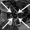
Image: T1 post-contrast subtraction imaging shows the mass having a black non-enhancing center, with a thin rim of peripheral enhancement.
Downloads
References
Vyas S, Markar S, Ezzat TM, Ajit A. Cystic lesions of the pancreas: current trends in approach and management. Postgrad Med J. 2011 Mar;87(1025):207-14.
Luchtrath H, Schriefers KH. [A pancreatic cyst with features of a so-called branchiogenic cyst]. Pathologe. 1985 Jul;6(4):217-9.
Adsay NV, Hasteh F, Cheng JD, Bejarano PA, Lauwers GY, Batts KP, et al. Lymphoepithelial cysts of the pancreas: a report of 12 cases and a review of the literature. Mod Pathol. 2002 May;15(5):492-501.
Truong LD, Rangdaeng S, Jordan PH, Jr. Lymphoepithelial cyst of the pancreas. Am J Surg Pathol. 1987 Nov;11(11):899-903.
Katz DS, Scatorchia GM, Wojtowycz AR, Botash RJ. Lymphoepithelial cyst of the pancreatic head. AJR Am J Roentgenol. 1995 Aug;165(2):489.
Mitchell ML. Fine needle aspiration biopsy of peripancreatic lymphoepithelial cysts. Acta Cytol. 1990 May-Jun;34(3):462-3.
Worrall NK, Drebin JA. Pancreaticoduodenectomy for lymphoepithelial cyst of the pancreas. Am Surg. 2000 Aug;66(8):732-4.
Kim WH, Lee JY, Park HS, Won HJ, Kim YH, Choi JY, et al. Lymphoepithelial cyst of the pancreas: comparison of CT findings with other pancreatic cystic lesions. Abdom Imaging. May 19.
Shinmura R, Gabata T, Matsui O. Lymphoepithelial cyst of the pancreas: case report with special reference to imaging--pathologic correlation. Abdom Imaging. 2006 Jan-Feb;31(1):106-9.
Zaheer A, Pokharel SS, Wolfgang C, Fishman EK, Horton KM. Incidentally detected cystic lesions of the pancreas on CT: review of literature and management suggestions. Abdom Imaging. Apr 26.
Nam SJ, Hwang HK, Kim H, Yu JS, Yoon DS, Chung JJ, et al. Lymphoepithelial cysts in the pancreas: MRI of two cases with emphasis of diffusion-weighted imaging characteristics. J Magn Reson Imaging. Sep;32(3):692-6.
Kudo D, Hashimoto N, Toyoki Y, Narumi S, Hakamada K. Usefulness of in-phase and out-of-phase magnetic resonance imaging for the detection of pancreatic lymphoepithelial cyst. Hepatogastroenterology. Jul-Aug;58(109):1403-5.
Ali S, Wilkinson N, Jensen CS, Gerke H. EUS-guided Trucut biopsies may enable the diagnosis of lymphoepithelial cysts of the pancreas. Report of two cases. JOP. 2009;10(4):409-12.
Allen PJ, D'Angelica M, Gonen M, Jaques DP, Coit DG, Jarnagin WR, et al. A selective approach to the resection of cystic lesions of the pancreas: results from 539 consecutive patients. Ann Surg. 2006 Oct;244(4):572-82.
Moparty B, Logrono R, Nealon WH, Waxman I, Raju GS, Pasricha PJ, et al. The role of endoscopic ultrasound and endoscopic ultrasound-guided fine-needle aspiration in distinguishing pancreatic cystic lesions. Diagn Cytopathol. 2007 Jan;35(1):18-25.
Jian B, Kimbrell HZ, Sepulveda A, Yu G. Lymphoepithelial cysts of the pancreas: endosonography-guided fine needle aspiration. Diagn Cytopathol. 2008 Sep;36(9):662-5.
Nasr J, Sanders M, Fasanella K, Khalid A, McGrath K. Lymphoepithelial cysts of the pancreas: an EUS case series. Gastrointest Endosc. 2008 Jul;68(1):170-3.
Karim Z, Walker B, Lam E. Lymphoepithelial cysts of the pancreas: the use of endoscopic ultrasound-guided fine-needle aspiration in diagnosis. Can J Gastroenterol. Jun;24(6):348-50.
Takamatsu S, Maruyama M, Sugano N, Ebuchi M. Lymphoepithelial cyst of the pancreas. Journal of Hepato-Biliary-Pancreatic Surgery. 1996;3(4):485-90.
Kaiserling E, Seitz KH, Rettenmaier G, Seidel W, Kahlfuss R, Walz-Mattmuller R, et al. Lymphoepithelial cyst of the pancreas. Clinical, morphological, and immunohistochemical findings. Zentralbl Pathol. 1991;137(5):431-8.
Hamamoto I, Ishimura K, Okada S, Kobayashi S, Kushida Y, Maeba T, et al. Extrapancreatic lymphoepithelial cyst of the pancreas. Journal of Hepato-Biliary-Pancreatic Surgery. 1996;3(2):186-91.
Misonou J, Iwabuchi K, Aizawa M, Murakami S. Mucoepidermoid carcinoma arising in a lymphoepithelial cyst--report of a case and review of the literature on branchiogenic carcinoma. Jpn J Surg. 1989 Jul;19(4):474-9.
Copyright (c) 2014 Srinivas Kavuturu, Nabeel E Sarwani, Fransesca M Ruggeiro, Isabelle Deshaies, Eric T Kimchi, Jussuf T Kaifi, Kevin F Staveley-O'Carroll, Niraj J Gusani

This work is licensed under a Creative Commons Attribution 4.0 International License.
As a member of Publisher International Linking Association, PILA, iMedPub Group’s JOP follows the Creative Commons Attribution License and Scholars Open Access publishing policies. Journal of the Pancreas is the Council Contributor Member of Council of Science Editors (CSE) and following the CSE slogan Education, Ethics, and Evidence for Editors.