
A First Report of Endoscopic Ultrasound for the Diagnosis of Pancreatic Amyloid Deposition in Immunoglobulin Light Chain (AL) Amyloidosis (Primary Amyloidosis)
Abstract
Context Pancreatic involvement in systemic light chain (AL)-amyloidosis is exceedingly rare. Prior reports of endoscopic ultrasound (EUS) for the diagnosis of amyloidosis are also limited. Case report We report the first description of EUS-guided fine needle aspiration (FNA) for the diagnosis of primary AL-amyloidosis involving the pancreas. Conclusion: EUS-FNA can be effectively utilized for the characterization and cytologic diagnosis of pancreatic amyloidosis and potentially other accessible extraluminal amyloid deposits.
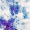
Image: Abundant amorphous acellular waxy appearing proteinaceous deposits.
Downloads
References
Ebert EC, Nagar M. Gastrointestinal manifestations of amyloidosis. Am J Gastroenterol 2008;103:776-87. [PMID: 18076735].
Kyle RA, Gertz MA. Primary systemic amyloidosis: clinical and laboratory features in 474 cases. Semin Hematol 1995;32:45-59. [PMID: 7878478].
Troussard X, Hurault de Ligny B, Gallet B, Ganeval D, Mandard JC, Ryckelynck JP, Leporrier M. Massive systemic amyloidosis associated with light chain deposition disease. Nephron 1989;52:139-43. [PMID: 2500613].
Onur MR, Yalniz M, Poyraz AK, Ozercan IH, Ozkan Y. Pancreatic islet cell amyloidosis manifesting as a large pancreas. Korean J Radiol 2012;13:94-7. [PMID: 22247642].
Gandolfi L, Colecchia A, Leo P, Caletti G, Rossi A, Primerano A, Torresan F. Endoscopic ultrasongraphy in the diagnosis of gastrointestinal amyloid deposits: clinical case report. Endoscopy 1995;27:132-4. [PMID: 7601026].
Grape T, Wurm Johansson G, Eriksson M, Toth E, Thorlacius H. Primary gastroduodenal amyloidosis. Endoscopy 2011;43:E288. [PMID: 21915830].
Shuttleworth E, Keld R, Willert R, Benbow EW. Amyloidosis: an EUS view. Gastrointest Endosc 2012;75:218-20. [PMID: 21470605].
Sawada T, Adachi Y, Akino K, Arimura Y, Ishida T, Ishii Y, Endo T. Endoscopic features of primary amyloidosis of the stomach. Endoscopy 2012;44:E275-6. [PMID: 22814919].
Halliday BE, Silverman JF, Finley JL. Fine-needle aspiration cytology of amyloid associated with nonneoplastic and malignant lesions. Diagn Cytopathol 1998;18:270-5. [PMID: 9557261].
Michael CW, Naylor B. Amyloid in cytologic specimens. Differential diagnosis and diagnostic pitfalls. Acta Cytol 1999;43:746-55. [PMID: 10518125].
Sahoo S, Reeves W, Demay RM. Amyloid tumor: a clinical and cytomorphologic study. Diagn Cytopathol 2003;28:325-8. [PMID: 12768639].
Copyright (c) 2014 Somashekar G Krishna, Manoop S Bhutani, Charles H Mosher, Gregg A Staerkel, Brian R Weston

This work is licensed under a Creative Commons Attribution 4.0 International License.
As a member of Publisher International Linking Association, PILA, iMedPub Group’s JOP follows the Creative Commons Attribution License and Scholars Open Access publishing policies. Journal of the Pancreas is the Council Contributor Member of Council of Science Editors (CSE) and following the CSE slogan Education, Ethics, and Evidence for Editors.