
Primary Squamous Cell Carcinoma of the Ampulla of Vater
Abstract
Context Squamous carcinoma of the ampulla of Vater is a very rare tumor with only three cases been reported so far. Case report Here, we report the case of a 68-year-old man who presented with painless obstructive jaundice, general fatigue, loss of appetite and weight loss. Laboratory tests revealed hypochromic anemia. Total and direct bilirubin, alkaline phosphatase, liver enzymes, carbohydrate antigen 19-9 (CA 19-9) and carcinoembryonic antigen (CEA) were all elevated. Abdominal ultrasonography and computed tomography showed a distended gallbladder, dilatation of the intra- and extra-hepatic bile ducts and enlargement of the pancreatic head. Endoscopic retrograde cholangiopancreatography revealed a bulging papilla with infiltrative growth at the ampulla of Vater but endoscopic biopsies were inconclusive. The patient was treated with classical Whipple’s pancreaticoduodenectomy. Histopathological examination showed a moderately differentiated squamous cell carcinoma. Multiple serial sectioning of the tumor specimen failed to detect an adenomatous component. Regional lymph nodes and resection margins were free of tumor and the disease was classified as stage IIA (T3N0M0) according to the TNM system. Adjuvant treatment was not given. Despite curative resection, multiple liver metastases developed after four months and the patient succumbed to progressive hepatic failure 5 months after the operation. Conclusion Primary pure squamous cell carcinoma of the ampulla of Vater is a very rare histological type of carcinoma. Clinical characteristics and optimal treatment are obscure. Primary surgical treatment with curative intent should be performed although this type of carcinoma associates with dismal prognosis.
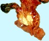
Image: Gray-white tumor tissue located within the wall of the ampulla of Vater.
Downloads
References
Pathak GS, Deshmukh SD, Yavalkar PA, Ashturkar AV. Coexistent ampullary squamous cell carcinoma with adenocarcinoma of the pancreatic duct. Saudi J Gastroenterol 2011; 17:411-3. [PMID 22064341]
Gupta A, Kumar S, Kumaresh TS, Gupta S, Makhija M, Singh B. Primary squamous cell carcinoma of the ampulla of Vater - a rare entity. The Internet Journal of Surgery 2010; 22(2).
Chen CM, Wu CS, Tasi SL, Hung CF, Chen TC. Squamous cell carcinoma of the ampulla of Vater: a case report. Changgeng Yi Xue Za Zhi 1996; 19:253-7. [PMID 8921644]
Ueno N, Sano T, Kanamaru T, Tanaka K, Nishihara T, Idei Y, Yamamoto M, Okuno T, Kawaguchi K. Adenosquamous cell carcinoma arising from the papilla major. Oncol Rep 2002; 9:317-20. [PMID 11836599]
Song HG, Yoo KS, Ju NR, Park JC, Jung JO, Shin WG, Moon JH, Kim JP, Kim KO, Park CH, Hahn T, Park SH, Kim JH, Lee IJ, Min SK, Park CK. A case of adenosquamous carcinoma of the papilla of Vater. Korean J Gastroenterol 2006; 48:132-6. [PMID 16929159]
Yavuz E, Kapran Y, Ozden I, Bulut T, Dizdaroğlu F. Pancreatobiliary adenosquamous carcinoma (report of two cases). Pathologica 2000; 92:323-6. [PMID 11198466]
Kimura W, Futakawa N, Zhao B. Neoplastic diseases of the papilla of Vater. J Hepatobiliary Pancreat Surg 2004; 11:223-31. [PMID 15368105]
Fischer HP, Zhou H. Pathogenesis of carcinoma of the papilla of Vater. J Hepatobiliary Pancreat Surg 2004; 11:301-9. [PMID 15549428]
Westgaard A, Pomianowska E, Clausen OP, Gladhaug IP. Intestinal-type and pancreatobiliary-type adenocarcinomas: how does ampullary carcinoma differ from other periampullary malignancies? Ann Surg Oncol 2013; 20:430-9. [PMID 22956064]
Hong SM, Kim MJ, Jang KT, Yoon GS, Cho H, Frierson HF, Yu E. Adenosquamous carcinoma of extrahepatic bile duct: clinicopathologic study of 12 cases. Int J Clin Exp Pathol 2008; 1:147-56. [PMID 18784802]
Yamana I, Kawamoto S, Nagao S, Yoshida T, Yamashita Y. Squamous cell carcinoma of the hilar bile duct. Case Rep Gastroenterol 2011; 5:463-70. [PMID 21960950]
Abbas R, Willis J, Kinsella T, Siegel C, Sanabria J. Primary squamous cell carcinoma of the main hepatic bile duct. Can J Surg 2008; 51:E85-6. [PMID 18815636]
Lim SH, Yang HW, Kim A, Cha SW, Jung SH, Go H, Lee WC. Adenosquamous carcinoma of extrahepatic bile duct: a case report. Korean J Intern Med 2007; 22:206-10. [PMID 17939340]
Sugawara G, Yamaguchi A, Isogai M, Watanabe Y, Kaneoka Y, Suzuki M. Small cell neuroendocrine carcinoma of the ampulla of Vater with foci of squamous differentiation: a case report. J Hepatobiliary Pancreat Surg 2004; 11:56-60. [PMID 15747032]
Kodavatiganti R, Campbell F, Hashmi A, Gollins SW. Primary squamous cell carcinoma of the pancreas: a case report and review of the literature. J Med Case Rep 2012; 6:295. [PMID 22973995]
Copyright (c) 2014 Helen Bolanaki, Alexandra Giatromanolaki, Efthimios Sivridis, Anastasios J Karayiannakis

This work is licensed under a Creative Commons Attribution 4.0 International License.
As a member of Publisher International Linking Association, PILA, iMedPub Group’s JOP follows the Creative Commons Attribution License and Scholars Open Access publishing policies. Journal of the Pancreas is the Council Contributor Member of Council of Science Editors (CSE) and following the CSE slogan Education, Ethics, and Evidence for Editors.