
Subcutaneously Inoculated Cells and Implanted Pancreatic Cancer Tissue Show Different Patterns of Metastases in Syrian Golden Hamsters
Abstract
Context We studied behavior of the subcutaneously implanted pancreatic tumors and the process of metastasis using syngeneic Syrian golden hamsters. Design HaP-T1, a cell line derived from nitrosamine-induced pancreatic cancer in Syrian golden hamsters was used for this experiment. Thirty-five animals were divided into two groups: subcutaneous cell inoculation and subcutaneous tissue implantation. The tumor tissue was obtained from subcutaneously implanted cancer cells. One month after implantation, the tumors were resected and studied histopathologically. The animals were followed-up weekly by palpation of the peripheral lymph nodes in order to identify local recurrence. After death, necropsy was performed. Liver, lungs and pancreas specimens were taken for histopathogical study and detection of K-ras point mutation using the PCR/RFLP method. Results The mean survival time in the subcutaneous cell inoculation group was 151±17.5 days, and in the subcutaneous tissue implantation group was 137±12.9 days. During the follow-up, 13 subcutaneously cell inoculated hamsters (86.7%) had right axillary lymph node metastasis while subcutaneously tissue implanted hamsters did not show any palpable lymph nodes. After necropsy, 10 of the 20 subcutaneously tissue implanted animals (50%) showed metastases in the lungs at the histopathological level. However, 16 of the 20 subcutaneously tissue implantated animals (80%) showed K-ras point mutation in the lung specimens. The lungs of the animals of the subcutaneous cell inoculation group did not show any metastases. No metastases were found in the liver or the pancreas in either group. Conclusion This study suggests that homologous subcutaneous cell inoculation and subcutaneous tissue implantation models showed completely different patterns of metastasis. These models may aid further research to clarify the mechanisms of metastasis in pancreatic cancer.
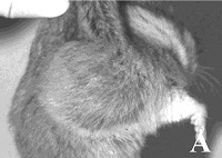
Image: Appearance of the right lymph node in a case from the SCI group.
Downloads
References
Nicolson GL, Poste G. Tumor implantation and invasion at metastatic sites. Int Rev Exp Pathol 1983; 25:77-181.
Pour PM, Wilson RB. Experimental tumors of the pancreas. In: Moossa AR, ed. Tumor of Pancreas. 1st ed. Baltimore: Waverly Press, 1980: 37-158.
Saito S, Nishimura N, Kubota Y, Yamazaki K, Shibuya T, Sasaki H. Establishment and characterization of a cultured cell line derived from nitrosamine-induced pancreatic ductal adenocarcinoma in Syrian golden hamsters. Gastroenterol Jpn 1988; 23:183-94.
Morioka CY, Saito S, Ohzawa K, Watanabe A. Homologous orthotopic implantation models of pancreatic ductal cancer in Syrian golden hamsters: which is better for metastasis research-cell implantation or tissue implantation? Pancreas 2000; 20:152-7.
Altman DG. Practical statistics for medical research. 1st ed. New York: Chapman and Hall, CRC, 1999.
Hanna N, Fidler IJ. Expression of metastatic potential of allogenic and xenogeneic neoplasms in young nude mice. Cancer Res 1981; 41:438-44.
Klein JR, Bevan MJ. Secretion of immune interferon and generation of cytotoxic T cell activity in nude mice are dependent on interleukin 2: age-associated endogenous production of interleukin 2 in nude mice. J Immunol 1983; 130:1780-3.
Kyriazis AP, DiPersio L, Michael GJ, Pesce AJ, Stinnett JD. Growth patterns and metastatic behavior of human tumors growing in athymic mice. Cancer Res 1978; 38:3186-90.
Kyriaziz AP, Kyriaziz AA, McCombs WB 3rd, Kereiakes JA. Biological behavior of human malignant tumors grown in the nude mouse. Cancer Res 1981; 41:3995-4000.
Fidler IJ. Origin and biology of cancer metastasis. Cytometry 1989; 10:673-80.
Greene HSN, Harvey EK. The relationship between the dissemination of tumor cells and the distribution of metastases. Cancer Res 1964; 24:799-811.
Hart IR. "Seed and soil" revisited mechanisms of site specific metastasis. Cancer Metastasis Rev 1982; 1:5-16.
Fidler IJ. Critical factors in the biology of human cancer metastasis: twenty-eight G.H.A. Clowes memorial award lecture. Cancer Res 1990; 6130-8.
Sakorafas GH, Tsiotou AG, Tsiotos GG. Molecular biology of pancreatic cancer; oncogenes, tumour suppressor genes, growth factors, and their receptors from a clinical perspective. Cancer Treat Rev 2000; 26:29-52.
Erill N, Cuatrecasas M, Sancho FJ, Farre A, Pour PM, Lluis F, Capella G. K-ras and p53 mutations in hamster pancreatic ductal adenocarcinomas and cell lines. Am J Pathol 1996; 149:1333-9.
Sugio K, Gazdar AF, Albores-Saavedra J, Kokkinakis DM. High yields of K-ras mutations in intraductal papillary mucinous tumors and invasive adenocarcinomas induced by N-nitroso(2-hydroxypropyl)(2-oxopropyl)amine in the pancreas of female Syrian hamsters. Carcinogenesis 1996; 17:303-9.
Konishi Y, Tsutsumi M, Tsujiuchi T. Mechanistic analysis of pancreatic ductal carcinogenesis in hamsters. Pancreas 1998; 16:300-6.
Copyright (c) 2000 Cintia Yoko Morioka, Seiji Saito, Kouji Ohzawa, Shinji Asano, Yasuhide Hibino, Yuji Nakada, Kei-ichiro Kita, Akiharu Watanabe

This work is licensed under a Creative Commons Attribution 4.0 International License.
As a member of Publisher International Linking Association, PILA, iMedPub Group’s JOP follows the Creative Commons Attribution License and Scholars Open Access publishing policies. Journal of the Pancreas is the Council Contributor Member of Council of Science Editors (CSE) and following the CSE slogan Education, Ethics, and Evidence for Editors.