
Pancreatic Schwannoma: A Case Report and Literature Review with Special Reference to Imaging Features
Abstract
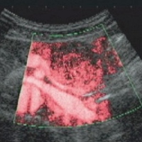
Context We report the imaging features of pancreatic schwannomas, a rare benign type of pancreatic tumor. Case report A 66-year-old woman was admitted to our hospital with a pancreatic tumor indicated in medical examinations. Computed tomography (CT), magnetic resonance imaging (MRI) and endoscopic ultrasonography (EUS) revealed a solid and cystic tumor, 3 cm in diameter, within the body of the pancreas. Contrast-enhanced CT, MRI and ultrasonography showed partial enhancement in the solid component. Endoscopic retrograde cholangiopancreatography (ERCP) and angiography showed no abnormal findings. A distal pancreatectomy together with a splenectomy and lymph node dissection were performed with a tentative diagnosis of mucinous cystic neoplasm of the pancreas. The cut surface of the resected pancreas showed a well-demarcated, pale yellow, solid tumor within the pancreas parenchyma. Histopathological examination of the tumor revealed proliferation of the spindle cells showing interlacing and palisading patterns. Immunohistochemically, these spindle cells were positive for S-100 protein and vimentin, and negative for alpha-smooth muscle actin, CD34, and cytokeratin. Thus the tumor was diagnosed as a pancreatic schwannoma. Conclusion CT and US can detect pancreatic schwannomas as solid and cystic masses, and MRI shows a relatively characteristic feature. Imaging procedures such as CT, MRI and US are able to differentiate a pancreatic tumor, such as a pancreatic schwannoma.
Image: Contrast-enhanced US shows partial enhancement at the solid component.
Downloads
References
Ferrozzi F, Bova D, Garlaschi G. Pancreatic schwannoma: report of three cases. Clin Radiol 1995; 50:492-5. [PMID 7614798] (FULL TEXT: http://www.sciencedirect.com/science?_ob=MImg&_imagekey=B6WCP-4JXR7NM-D-1&_cdi=6744&_user=839424&_orig=browse&_coverDate=07%2F31%2F1995&_sk=999499992&view=c&wchp=dGLbVlz-zSkzk&md5=0e72280eb20ed4121d2a959009959fcf&ie=/sdarticle.pdf)
Paranjape C, Johnson SR, Khwaja K, Goldman H, Kruskal JB, Hanto DW. Clinical characteristics, treatment, and outcome of pancreatic schwannoma. J Gastrointest Surg 2004; 8:706-12. [PMID 15358332] (FULL TEXT: http://www.springerlink.com/content/u7440255384081x6/fulltext.pdf)
Kim SH, Choi BI, Han MC, Kim YI. Retroperitoneal neurilemoma: CT and MRI findings. AJR Am J Roentgenol 1992; 159:1023-6. [PMID 1414767] (FULL TEXT: http://www.ajronline.org/cgi/reprint/159/5/1023)
Feldman L, Philpotts LE, Reinhold C, Duguid WP, Rosenberg L. Pancreatic schwannoma: report of two cases and review of the literature. Pancreas 1997; 15:99-105. [PMID 9211499] (FULL TEXT: http://journals.lww.com/pancreasjournal/pages/articleviewer.aspx?year=1997&issue=07000&article=00014&type=abstract)
Soumaoro LT, Teramoto K, Kawamura T, Nakamura N, Sanada T, Sugihara K, Arii S. Benign schwannoma of the pancreas. J Gastrointest Surg 2005; 9:288-90. [PMID 15694826] (FULL TEXT: http://www.springerlink.com/content/9275170233356625/fulltext.pdf)
Okuma T, Hirota M, Nitta H, Saito S, Yagi T, Ida S, et al. Pancreatic schwannoma: Report of a case. Surg Today 2008; 38:266-70. [PMID 18307004] (FULL TEXT: http://www.springerlink.com/content/kx6244q3224h0706/fulltext.pdf)
Verocay J. Zur Kenntnis der "Neurofibrome". Beitr Pathol Anat Allg Pathol 1910; 48:1-69.
Tomozawa S, Masaki T, Matsuda K, Yokoyama T, Ishida T, Muto T. A schwannoma of the cecum: case report and review of Japanese schwannomas in the large intestine. J Gastroenterol 1998; 33:872-5. [PMID 9853563] (FULL TEXT: http://www.springerlink.com/content/6bpvycy6ewq0uejl/fulltext.pdf)
Morita S, Okuda J, Sumiyoshi K, Taketani M, Moriguchi A, Katsu K, Tanigawa N. Pancreatic schwannoma: report of a case. Surg Today 1999; 29:1093-7. [PMID 10554337] (FULL TEXT: http://www.springerlink.com/content/ce4pq8nqg2yqkbbj/fulltext.pdf)
Hsaio WC, Lin PW, Chang KC. Benign retroperitoneal Schwannoma mimicking a pancreatic cystic tumor: case report and literature review. Hepatogastroenterology 1998; 45:2418-20. [PMID 9951935]
Novellas S, Chevallier P, Saint Paul MC, Gugenheim J, Bruneton JN. MRI features of a pancreatic schwannoma. Clin Imaging 2005; 29:434-6. [PMID 16274826] (FULL TEXT: http://www.sciencedirect.com/science?_ob=ArticleURL&_udi=B6T5C-4HGMPKC-C&_user=839424&_rdoc=1&_fmt=&_orig=search&_sort=d&_docanchor=&view=c&_acct=C000045367&_version=1&_urlVersion=0&_userid=839424&md5=26f2db451760069e822567ca07025668)
Wu YL, Yan HC, Chen LR, Chen J, Gao SL, Li JT. Pancreatic benign schwannoma treated by simple enucleation: case report and review of literature. Pancreas 2005; 31:286-8. [PMID 16163062] (FULL TEXT: http://journals.lww.com/pancreasjournal/pages/articleviewer.aspx?year=2005&issue=10000&article=00013&type=abstract)
Bui TD, Nguyen T, Huerta S, Gu M, Hsiang D. Pancreatic schwannoma: a case report and review of the literature. JOP. J Pancreas (Online) 2004; 5:520-6. [PMID 15536295] (FULL TEXT: http://www.joplink.net/prev/200411/14.html)
Yu Rs, Sun JZ. Pancreatic schwannoma: CT findings. Abdom Imaging 2006; 31:103-5. [PMID 16132429] (FULL TEXT: http://www.springerlink.com/content/r2n586u3h322n834/fulltext.html)
Tan G, Vitellas K, Morrison C, Frankel WL. Cystic schwannoma of the pancreas. Ann Diagn Pathol 2003; 7:285-91. [PMID 14571430] (FULL TEXT: http://www.sciencedirect.com/science?_ob=ArticleURL&_udi=B6W9Y-49PR9CB-7&_user=839424&_rdoc=1&_fmt=&_orig=search&_sort=d&_docanchor=&view=c&_acct=C000045367&_version=1&_urlVersion=0&_userid=839424&md5=a5755ea9e8e97fe1111e6828c99280b4)
Coombs RJ. Case of the season. Malignant neurogenic tumor of duodenum and pancreas. Semin Roentgenol 1990; 25:127-9. [PMID 2112269] (FULL TEXT: http://www.sciencedirect.com/science?_ob=ArticleURL&_udi=B75KX-4FS0Y96-7D&_user=606145&_coverDate=04%2F30%2F1990&_rdoc=2&_fmt=high&_orig=browse&_srch=doc-info%28%23toc%2313190%231990%23999749997%23586702%23FLP%23display%23Volume%29&_cdi=13190&_sort=d&_docanchor=&_ct=7&_acct=C000031398&_version=1&_urlVersion=0&_userid=606145&md5=028f4ff43f44cb6a72038cd0eacc9ab3)
Walsh MM, Brandspigel K. Gastrointestinal bleeding due to pancreatic schwannoma complicating von Recklinghausen's disease. Gastroenterology 1989; 97:1550-1. [PMID 2511055] (FULL TEXT: http://download.journals.elsevierhealth.com/pdfs/journals/0016-5085/PII0016508589904022.pdf)
Eggermont A, Vuzevski V, Huisman M, De Jong K, Jeekel J. Solitary malignant schwannoma of the pancreas: report of a case and ultrastructural examination. J Surg Oncol 1987; 36:21-5. [PMID 3626558] (FULL TEXT: http://www3.interscience.wiley.com/cgi-bin/fulltext/112719569/PDFSTART)
Moller Pedersen V, Hede A, Graem N. A solitary malignant schwannoma mimicking a pancreatic pseudocyst. A case report. Acta Chir Scand 1982; 148:697-8. [PMID 7170905]
Lee JS, Kim HS, Jung JJ, Han SW, Kim YB. Ancient schwannoma of the pancreas mimicking a cystic tumor. Virchows Arch 2001; 439:697-9. [PMID 11764392] (FULL TEXT: http://www.springerlink.com/content/3xb1rl2n1k8pgu8t/fulltext.html)
Almo KM, Traverso LW. Pancreatic schwannoma: an uncommon but important entity. J Gastrointest Surg 2001; 5:359-63. [PMID 11985975] (FULL TEXT: http://www.springerlink.com/content/u242458885443173/fulltext.pdf)
Brown SZ, Owen DA, O'Connell JX, Scudamore CH. Schwannoma of the pancreas: a report of two cases and a review of the literature. Mod Pathol 1998; 11:1178-82. [PMID 9872648]
Todd KE, Lewis MP, Gloor B, Kusske AM, Ashley SW, Reber HA. Management decisions for unusual periampullary tumors. Am Surg 1997; 63:927-32. [PMID 9322675]
Sugiyama M, Kimura W, Kuroda A, Muto T. Schwannoma arising from peripancreatic nerve plexus. AJR Am J Roentgenol 1995; 165:232. [PMID 7785620] (FULL TEXT: http://www.ajronline.org/cgi/reprint/165/1/227.pdf)
Steven K, Burcharth F, Holm N, Pedersen IK. Single stage pancreaticoduodenectomy(Whipple's procedure), radical cystectomy and bladder substitution with the urethral Kock reservoir. Case report. Scand J Urol Nephrol 1994; 28:199-200. [PMID 7939474]
Melato M, Bucconi S, Marus W, Spivach A, Perulli A, Mucelli RP. The schwannoma: an uncommon type of cystic lesion of the pancreas. Ital J Gastroenterol 1993; 25:385-7. [PMID 8280901]
David S, Barkin JS. Pancreatic schwannoma. Pancreas 1993; 8:274-6. [PMID 8460103] (FULL TEXT: http://journals.lww.com/pancreasjournal/Citation/1993/03000/Pancreatic_Schwannoma.22.aspx)
Urban BA, Fishman EK, Hruban RH, Cameron JL. CT findings in cystic schwannoma of the pancreas. J Comput Assist Tomogr 1992; 16:492-3. [PMID 1592939] (FULL TEXT: http://journals.lww.com/jcat/Citation/1992/05000/CT_Findings_in_Cystic_Schwannoma_of_the_Pancreas.30.aspx)
Liessi G, Barbazza R, Sartori F, Sabbadin P, Scapinello A. CT and MR imaging of melanocytic schwannomas; report of three cases. Eur J Radiol 1990; 11:138-42. [PMID 2253635] (FULL TEXT: http://www.sciencedirect.com/science?_ob=MImg&_imagekey=B6T6F-4CDJ1DB-D-1&_cdi=5029&_user=839424&_orig=search&_coverDate=10%2F31%2F1990&_sk=999889997&view=c&wchp=dGLbVlz-zSkzV&md5=44c64ef4a2acf29b1f7760eca5b18b57&ie=/sdarticle.pdf)
Hirabayashi K, Yasuda M, Uemura S, Itoh H, Itoh J, Yazawa N, et al. Cytological features of the cystic fluid of pancreatic schwannoma with cystic degeneration. A case report. JOP. J Pancreas (Online) 2008; 9:203-8. [PMID 18326930] (FULL TEXT: http://www.joplink.net/prev/200803/11.html)
Enzinger FM, Weiss SW. Benign tumors of the peripheral nerves. In: Enzinger FM ed. Soft Tissue Tumors, 3rd ed. St Louis:Mosby, 1995:821-8.
Copyright (c) 2010 Shiji Suzuki, Satoshi Kaji, Nobusada Koike, Nobuhiko Harada, Tsuneo Hayashi, Mamoru Suzuki, Fujio Hanyu, Shinichi Ban

This work is licensed under a Creative Commons Attribution 4.0 International License.
As a member of Publisher International Linking Association, PILA, iMedPub Group’s JOP follows the Creative Commons Attribution License and Scholars Open Access publishing policies. Journal of the Pancreas is the Council Contributor Member of Council of Science Editors (CSE) and following the CSE slogan Education, Ethics, and Evidence for Editors.