
Intraductal Oncocytic Papillary Neoplasm of the Pancreas: A Radio-Pathological Case Study
Abstract
Context An intraductal oncocytic papillary neoplasm is a rare pancreatic tumor with the potential of developing invasive carcinoma. Its differentiation from other cystic-like neoplasms of the pancreas, such as intraductal papillary mucinous neoplasms, is a challenge for pancreatic imaging. Case report We present the case of a 76-year-old male with painless jaundice caused by an intraductal oncocytic papillary neoplasm of the pancreas. The imaging findings on computed tomography, magnetic resonance including diffusion-weighted imaging, and 18F-fluorodeoxyglucose positron emission tomography are presented and the radio-pathological correlations are discussed. Conclusion An intraductal oncocytic papillary neoplasm of the pancreas appears as a cystic tumor communicating with the dilated pancreatic duct featuring intraductal tumor nodules. Intraductal oncocytic papillary neoplasms show a high 18F-fluorodeoxyglucose-uptake in positron emission tomography and low diffusion values in diffusion-weighted imaging including apparent diffusion coefficient maps which may be a valuable attribute in distinguishing these rare lesions from intraductal papillary mucinous neoplasms.
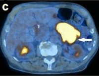
Image: Original attenuation-corrected coronal PET image.
Downloads
References
Adsay NV, Adair CF, Heffess CS, Klimstra DS. Intraductal oncocytic papillary neoplasms of the pancreas. Am J Surg Pathol 1996; 20:980-94. [PMID 8712298] (FULL TEXT: http://journals.lww.com/ajsp/pages/articleviewer.aspx?year=1996&issue=08000&article=00007&type=abstract)
Jyotheeswaran S, Zotalis G, Penmetsa P, Levea CM, Schoeniger LO, Shah AN. A newly recognized entity: intraductal "oncocytic" papillary neoplasm of the pancreas. Am J Gastroenterol 1998; 93:2539-43. [PMID 9860422] (FULL TEXT: http://www.nature.com/ajg/journal/v93/n12/full/ajg1998591a.html)
Nobukawa B, Suda K, Suyama M, Ariyama J, Beppu T, Futagawa S. Intraductal oncocytic papillary carcinoma with invasion arising from the accessory pancreatic duct. Gastrointest Endosc 1999; 50:864-6. [PMID 10570358] (FULL TEXT: http://www.giejournal.org/article/S0016-5107(99)70180-X/fulltext)
Noji T, Kondo S, Hirano S, Ambo Y, Tanaka E, Katoh C, et al. Intraductal oncocytic papillary neoplasm of the pancreas shows strong positivity on FDG-PET. Int J Gastrointest Cancer 2002; 32:43-6. [PMID 12630769] (FULL TEXT: http://www.springerlink.com/content/4002tj83v411h47u/fulltext.pdf)
Oku T, Maeda M, Wada Y, Waga E, Ono K, Nagamachi Y, et al. Intraductal oncocytic papillary neoplasm having clinical characteristics of mucinous cystic neoplasm and a benign histology. JOP. J Pancreas (Online) 2007; 8:206-13. [PMID 17356245] (FULL TEXT: http://www.joplink.net/prev/200703/06.html)
Shima Y, Yagi T, Inagaki M, Sadamori H, Tanaka N, Horimi T, et al. Intraductal oncocytic papillary neoplasm of the pancreas with celiac artery compression syndrome and a jejunal artery aneurysm: report of a case. Surg Today 2005; 35:86-90. [PMID 15622472] (FULL TEXT: http://www.springerlink.com/content/q2w37lku3jwqxq9g/fulltext.pdf)
Thompson K, Castelli MJ, Gattuso P. Metastatic papillary oncocytic carcinoma of the pancreas to the liver diagnosed by fine-needle aspiration. Diagn Cytopathol 1998; 18:291-6. [PMID 9557266] (FULL TEXT: http://www3.interscience.wiley.com/cgi-bin/fulltext/39163/PDFSTART)
Kato Y, Nakagouri T, Konishi M, Takahashi S, Gotoda N, Hasebe T, Kinosita T. Intraductal oncocytic papillary neoplasm of the pancreas with strong accumulation on FDG-PET. Hepatogastroenterology 2008; 55:900-2. [PMID 18705293]
Ishida M, Egawa S, Aoki T, Sakata N, Mikami Y, Motoi F, et al. Characteristic clinicopathological features of the types of intraductal papillary-mucinous neoplasms of the pancreas. Pancreas 2007; 35:348-52. [PMID 18090241] (FULL TEXT: https://journals.lww.com/pages/login.aspx?ReturnUrl=%2fpancreasjournal%2fsecure%2fpages%2fpurchase.aspx%3fan%3d00006676-200711000-00009)
Patel SA, Adams R, Goldstein M, Moskaluk CA. Genetic analysis of invasive carcinoma arising in intraductal oncocytic papillary neoplasm of the pancreas. Am J Surg Pathol 2002; 26:1071-7. [PMID 12170096] (FULL TEXT: http://journals.lww.com/ajsp/pages/articleviewer.aspx?year=2002&issue=08000&article=00014&type=abstract)
Hruban R, Pitman MB, Klimstra DS. AFIP Atlas of Tumor Pathology. Tumors of the Pancreas. Series 4, Fasc 6: Washington, American Registry of Pathology in collaboration with Armed Forces Institute of Pathology, 2007, pp 23-50.
Itai Y, Minami M. Intraductal papillary-mucinous tumor and mucinous cystic neoplasm: CT and MR findings. Int J Gastrointest Cancer 2001; 30:47-63. [PMID 12489580] (FULL TEXT: http://www.springerlink.com/content/f6k23232g4876178/fulltext.pdf)
Strobel K, Heinrich S, Bhure U, Soyka J, Veit-Haibach P, Pestalozzi BC, et al. Contrast-enhanced 18F-FDG PET/CT: 1-stop-shop imaging for assessing the resectability of pancreatic cancer. J Nucl Med 2008; 49:1408-13. [PMID 18703604] (FULL TEXT: http://jnm.snmjournals.org/cgi/content/full/49/9/1408)
Yoshioka M, Sato T, Furuya T, Shibata S, Andoh H, Asanuma Y, et al. Positron emission tomography with 2-deoxy-2-[(18)F] fluoro- d-glucose for diagnosis of intraductal papillary mucinous tumor of the pancreas with parenchymal invasion. J Gastroenterol 2003; 38:1189-93. [PMID 14714260] (FULL TEXT: http://www.springerlink.com/content/5j1lm8h61w81rfuc/fulltext.pdf)
Sperti C, Bissoli S, Pasquali C, Frison L, Liessi G, Chierichetti F, Pedrazzoli S. 18-fluorodeoxyglucose positron emission tomography enhances computed tomography diagnosis of malignant intraductal papillary mucinous neoplasms of the pancreas. Ann Surg 2007; 246:932-7. [PMID 18043094] (FULL TEXT: http://journals.lww.com/annalsofsurgery/pages/articleviewer.aspx?year=2007&issue=12000&article=00004&type=abstract)
Blake MA, McKernan M, Setty B, Fischman AJ, Mueller PR. Renal oncocytoma displaying intense activity on 18F-FDG PET. AJR Am J Roentgenol 2006; 186:269-70. [PMID 16357422] (FULL TEXT: http://www.ajronline.org/cgi/content/full/186/1/269)
Kim DJ, Chung JJ, Ryu YH, Hong SW, Yu JS, Kim JH. Adrenocortical oncocytoma displaying intense activity on 18F-FDG-PET: a case report and a literature review. Ann Nucl Med 2008; 22:821-8. [PMID 19039562] (FULL TEXT: http://www.springerlink.com/content/165g1u00813754x1/fulltext.pdf)
Subramaniam RM, Durnick DK, Peller PJ. F-18 FDG PET/CT imaging of submandibular gland oncocytoma. Clin Nucl Med 2008; 33:472-4. [PMID 18580232] (FULL TEXT: http://journals.lww.com/nuclearmed/pages/articleviewer.aspx?year=2008&issue=07000&article=00005&type=abstract)
Hagino K, Tsunoda A, Ishihara A, Kishimoto S, Suzuki T, Hara A. Oncocytoma in the parotid gland presenting a remarkable increase in fluorodeoxyglucose uptake on positron emission tomography. Otolaryngol Head Neck Surg 2006; 134:708-9. [PMID 16564402] (FULL TEXT: http://www.otojournal.org/article/PIIS0194599805004031/fulltext)
Vilanova JC, Barcelo J. Diffusion-weighted whole-body MR screening. Eur J Radiol 2008; 67:440-7. [PMID 18430538] (FULL TEXT: http://www.ejradiology.com/article/PIIS0720048X08001733/fulltext)
Bruegel M, Rummeny EJ. Hepatic metastases: use of diffusion-weighted echo-planar imaging. Abdom Imaging 2009; May 27. [PMID 19471997] (FULL TEXT: http://www.springerlink.com/content/q0h7353082167771/fulltext.html)
Matsuki M, Inada Y, Nakai G, Tatsugami F, Tanikake M, Narabayashi I, et al. Diffusion-weighted MR imaging of pancreatic carcinoma. Abdom Imaging 2007; 32:481-3. [PMID 17431713] (FULL TEXT: http://www.springerlink.com/content/08714340n8032234/fulltext.html)
Takeuchi M, Matsuzaki K, Kubo H, Nishitani H. High-b-value diffusion-weighted magnetic resonance imaging of pancreatic cancer and mass-forming chronic pancreatitis: preliminary results. Acta Radiol 2008; 49:383-6. [PMID 18415779] (FULL TEXT: http://informahealthcare.com/doi/full/10.1080/02841850801895381)
Yamashita Y, Namimoto T, Mitsuzaki K, Urata J, Tsuchigame T, Takahashi M, Ogawa M. Mucin-producing tumor of the pancreas: diagnostic value of diffusion-weighted echo-planar MR imaging. Radiology 1998; 208:605-9. [PMID 9722835] (FULL TEXT: http://radiology.rsna.org/content/208/3/605.long)
Kartalis N, Lindholm TL, Aspelin P, Permert J, Albiin N. Diffusion-weighted magnetic resonance imaging of pancreas tumours. Eur Radiol 2009; 19:1981-90. [PMID 19308414] (FULL TEXT: http://www.springerlink.com/content/m3310p70g6121thw/fulltext.html)
Ozsunar Y, Sorensen AG. Diffusion- and perfusion-weighted magnetic resonance imaging in human acute ischemic stroke: technical considerations. Top Magn Reson Imaging 2000; 11:259-272. [PMID 11142625] (FULL TEXT: https://journals.lww.com/topicsinmri/secure/pages/purchase.aspx?an=00002142-200010000-00003)
Inan N, Arslan A, Akansel G, Anik Y, Demirci A. Diffusion-weighted imaging in the differential diagnosis of cystic lesions of the pancreas. AJR Am J Roentgenol 2008; 191:1115-21. [PMID 18806153] (FULL TEXT: http://www.ajronline.org/cgi/content/full/191/4/1115)
Naganawa S, Kawai H, Fukatsu H, Sakurai Y, Aoki I, Miura S, et al. Diffusion-weighted imaging of the liver: technical challenges and prospects for the future. Magn Reson Med Sci 2005; 4:175-186. [PMID 16543702] (FULL TEXT: http://www.jstage.jst.go.jp/article/mrms/4/4/175/_pdf)
Copyright (c) 2010 Michael A Fischer, Olivio Donati, Stefan Heinrich, Achim Weber, Thomas F Hany, Davide Soldini, Hatem Alkadhi, Borut Marincek, Hans Scheffel

This work is licensed under a Creative Commons Attribution 4.0 International License.
As a member of Publisher International Linking Association, PILA, iMedPub Group’s JOP follows the Creative Commons Attribution License and Scholars Open Access publishing policies. Journal of the Pancreas is the Council Contributor Member of Council of Science Editors (CSE) and following the CSE slogan Education, Ethics, and Evidence for Editors.