
Computed Tomography Scan
Abstract
The diagnosis of different pancreatic diseases has recently become a recurrent problem. In cases with pancreatic head mass the main question is the differentiation between malignant and benign lesions. When a neoplasm is suspected, the main task is to judge operability. The usefulness of computed tomography imaging in the evaluation of pancreatic carcinoma has been well established. In this article the authors discuss the possibilities of computed tomography (CT) in diagnostic work-up.
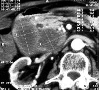
Image: CT after contrast medium administration shows multiple cystic lesions in the head of the pancreas.
Downloads
References
Baum U, Lell M, Nomayr A, Wolf H, Brunner T, Greess H, Bautz W. Mehrzeilen-Spiral-CT in der Diagnostik von Pankreastumoren. Radiologe 1999; 39:958-64.
Kim T, Murakami T, Takahashi S, Okada A, Hori M, Narumi Y, Nakamura H. Pancreatic CT imaging: effects of different injection rates and doses of contrast material. Radiology 1999; 212:219-25.
Richter GM, Wunsch C, Schneider B, Dux M, Klar E, Seelos R, Kauffmann GW. Hydro-CT in detection and staging of pancreatic carcinoma. Radiologe 1998; 38:279-86.
Freeny PC, Traverso LW, Ryan JA. Diagnosis and staging of pancreatic adenocarcinoma with dynamic computed tomography. Am J Surg 1993; 165:600-6.
Tabuchi T, Itoh K, Ohshio G, Kojima N, Maetani Y, Shibata T, Konishi J. Tumor staging of pancreatic adenocarcinoma using early- and late-phase helical CT. AJR Am J Roentgenol 1999; 173:375-80.
Barkin JS, Goldstein JA. Diagnostic approach to pancreatic cancer. Gastroenterol Clin North Am 1999; 28:709-22.
Bluemke DA, Cameron JL, Hruban RH, Pitt HA, Siegelman SS, Soyer P, Fishman EK. Potentially resectable pancreatic adenocarcinoma: spiral CT assessment with surgical and pathologic correlation. Radiology 1995; 197:381-5.
Sim JS, Choi BI, Han JK, Chung MJ, Chung JW, Park JH, Han MC. Helical CT anatomy of pancreatic arteries. Abdom Imaging 1996; 21:517-21.
O'Malley ME, Boland GW, Wood BJ, Fernandez-del Castillo C, Warshaw AL, Mueller PR. Adenocarcinoma of the head of the pancreas: determination of surgical unresectability with thin-section pancreatic-phase helical CT. AJR Am J Roentgenol 1999; 173:1513-8.
Yamada Y, Mori H, Kiyosue H, Matsumoto S, Hori Y, Maeda T. CT assessment of the inferior peripancreatic veins: clinical significance. AJR Am J Roentgenol 2000; 174:677-84.
Vedantham S, Lu D, Reber HA, Kadell B. Small peripancreatic veins: improved assessment in pancreatic cancer patients using thin-section pancreatic phase helical CT. AJR Am J Roentgenol 1998;170:377-83.
Irie H, Honda H, Kaneko K, Kuroiwa T, Yoshimitsu K, Masuda K. Comparison of helical CT and MR imaging in detecting and staging small pancreatic adenocarcinoma. Abdom Imaging 1997; 22:429-33.
Gorelick AB, Scheiman JM, Fendrick AM. Identification of patients with resectable pancreatic cancer: at what stage are we? Am J Gastroenterol 1998; 93:1995-6.
Hough TJ, Raptopoulos V, Siewert B, Matthews JB. Teardrop superior mesenteric vein: CT sign for unresectable carcinoma of the pancreas. AJR Am J Roentgenol 1999; 173:1509-12.
Ferrozzi F, Bova D, Campodonico F, De Chiara F, Passari A, Bassi P. Pancreatic metastases: CT assessment. Eur Radiol 1997; 7:241-5.
Ferrucci JT. Biliopancreatic malignancy current diagnostic possibilities: an overview. Ann Oncol 1999;10 (Suppl. 4):143-4.
Manes G, Kahl S, Glasbrenner B, Malfertheiner P. Chronic pancreatitis: diagnosis and staging. Ann Ital Chir 2000; 71:23-32.
Copyright (c) 2000 Tamás Winternitz, Hay Habib, Katalin Kiss, Tibor Tihanyi

This work is licensed under a Creative Commons Attribution 4.0 International License.
As a member of Publisher International Linking Association, PILA, iMedPub Group’s JOP follows the Creative Commons Attribution License and Scholars Open Access publishing policies. Journal of the Pancreas is the Council Contributor Member of Council of Science Editors (CSE) and following the CSE slogan Education, Ethics, and Evidence for Editors.