
Endoscopic Findings of Upper Gastrointestinal Lesions in Patients with Pancreatic Cancer
Abstract
Context Pancreatic cancer is often complicated with upper gastrointestinal lesions. However, there have been few endoscopic studies in pancreatic cancer patients. We retrospectively investigated the upper gastrointestinal lesions in patients with pancreatic cancer who underwent upper gastrointestinal endoscopy. Methods Upper gastrointestinal endoscopy was performed in 75 patients with pancreatic cancer between 2003 and 2010. We examined upper gastrointestinal lesions, such as gastroduodenal invasion, ulcers, esophagogastric varices, radiation-induced gastroduodenal mucosal lesions, and portal hypertensive gastropathy. Results Among the 53 patients with pancreatic cancer who underwent upper gastrointestinal endoscopy at diagnosis, 23 gastrointestinal lesions were observed in 20 patients (38%) as follows: gastroduodenal invasion (n=11), esophagogastric varices (n=7), gastroduodenal ulcers (n=3), portal hypertensive gastropathy (n=1) and duodenal metastasis (n=1). Among the 75 patients with pancreatic cancer, 56 gastrointestinal lesions were identified in 46 patients (61%) during the clinical course as follows: gastroduodenal invasion (n=20), esophagogastric varices (n=14), radiation-induced gastroduodenal mucosal lesions (n=9), gastroduodenal ulcers (except radiation-induced ulcers) (n=8), portal hypertensive gastropathy (n=3), duodenal metastasis (n=1), and gastrointestinal bleeding from unknown primary site (n=1). Twenty-nine (52%) of the 56 gastrointestinal lesions showed symptoms related to the lesions. Fifteen (27%) lesions were accompanied by upper gastrointestinal bleeding. Fourteen (25%) lesions developed according to the progression of pancreatic cancer. Conclusion We should pay attention to upper gastrointestinal lesions in patients with pancreatic cancer.
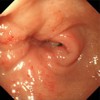
Image: Case of pancreatic head cancer with duodenal invasion.
Downloads
References
Cubilla A, Fitzgerald PJ. Pancreas cancer. I. Duct adenocarcinoma. A clinical-pathologic study of 380 patients. Pathol Annu 1978; 13 Pt 1: 241-89. [PMID 214741]
Sharon P, Stalnikovicz R, Rachmilewitz D. Endoscopic diagnosis of duodenal neoplasms causing upper gastrointestinal bleeding. J Clin Gastroenterol 1982; 4: 35-8. [PMID 6978900]
Lee P, Sutherland D, Feller ER. Massive gastrointestinal bleeding as the initial manifestation of pancreatic carcinoma. Int J Pancreatol 1994; 15: 223-7. [PMID 7930783]
Mao C, Domenico DR, Kim K, Hanson DJ, Howard JM. Observations on the developmental patterns and the consequences of pancreatic exocrine adenocarcinoma. Findings of 154 autopsies. Arch Surg 1995; 130: 125-34. [PMID 7848081]
Tio TL, Kimmings N, Rauws E, Jansen P, Tytgat G. Endosonography of gastroesophageal varices: evaluation and follow-up of 76 cases. Gastrointest Endosc 1995; 42: 145-50. [PMID 7590050]
Tomita H, Osada S, Matsuo M, Shimokawa K. Pancreatic cancer presenting with hematemesis from directly invading the duodenum: report of an unusual manifestation and review. Am Surg 2006; 72: 363-6. [PMID 16676866]
Mullan FJ, McKelvey ST. Pancreatic carcinoma presenting as bleeding from segmental gastric varices: pitfalls in diagnosis. Postgrad Med J 1990; 66: 401-3. [PMID 2371194]
Smith TA, Brand EJ. Pancreatic cancer presenting as bleeding gastric varices. J Clin Gastroenterol 2001; 32: 444-7. [PMID 11319321]
Shirato I, Nakamura S, Mitsunaga A, Shiratori K. Clinical features of esophago-gastric varices caused by pancreatic cancer (in Japanese with English abstract). Nippon Shokakibyo Gakkai Zasshi (JJSG) 2008; 105: 1186-92. [PMID 18678994]
Sobin LH, Christian W. UICC. TNM classification of malignant tumours. 6th ed. New York, Wiley-Liss, 2002.
Tajiri T, Yoshida H, Obara K, et al. General rules for recording endoscopic findings of esophagogastric varices (2nd edition). Dig Endosc 2010; 22: 1-9. [PMID 20078657]
Turrill FL, Mikkelsen WP. ‘’Sinistral’’ (left-sided) extrahepatic portal hypertension. Arch Surg 1969; 99: 365-8. [PMID 5306330]
Okusaka T, Ito Y, Ueno H, et al. Phase II study of radiotherapy combined with gemcitabine for locally advanced pancreatic cancer. Br J Cancer 2004; 91: 673-7. [PMID 15226765]
Wilkowski R, Thoma M, Bruns C, Wagner A, Heinemann V. Chemoradiotherapy with gemcitabine and continuous 5-FU in patients with primary inoperable pancreatic cancer. JOP 2006; 7: 349-60. [PMID 16832132]
Burris HA 3rd, Moore MJ, Andersen J, et al. Improvements in survival and clinical benefit with gemcitabine as first-line therapy for patients with advanced pancreas cancer: a randomized trial. J Clin Oncol 1997; 15: 2403-13. [PMID 9196156]
Ueno H, Okusaka T, Ikeda M, Takezako Y, Morizane C. An early phase II study of S-1 in patients with metastatic pancreatic cancer. Oncology 2005; 68: 171-8. [PMID 16006754]
Copyright (c) 2014 Koushiro Ohtsubo, Hiroyuki Watanabe, Hisatsugu Mouri, Kaname Yamashita, Kazuo Yasumoto, Seiji Yano

This work is licensed under a Creative Commons Attribution 4.0 International License.
As a member of Publisher International Linking Association, PILA, iMedPub Group’s JOP follows the Creative Commons Attribution License and Scholars Open Access publishing policies. Journal of the Pancreas is the Council Contributor Member of Council of Science Editors (CSE) and following the CSE slogan Education, Ethics, and Evidence for Editors.