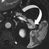
Intrapancreatic Accessory Spleen: The Usefulness of Arciform Arterial Enhancement for non-Invasive Diagnosis
Abstract
No abstract available.
Image: Arciform arterial enhancement at axial arterial-phase contrast enhanced MRI.
Downloads
References
Krishna SG, Heif MM, Sharma SG, Pandey T, Rego RF. Intrapancreatic accessory spleen: investigative dilemmas and role of EUS-guided FNA for diagnostic confirmation. JOP. J Pancreas (Online) 2011; 12(6):603-6.
Mortele KJ, Mortele B, Silverman SG. CT features of the accessory spleen. AJR Am J Roentgenol 2004; 183: 1653-1657.
Kim SH, Lee JM, Han JK, et al. Intrapancreatic accessory spleen: Findings on MR imaging, CT, US and scintigraphy, and the pathologic analysis. Korean J Radiol 2008; 9: 162-174.
Low G, Panu A, Millo N, Leen E. Multimodality imaging of neoplastic and nonneoplastic solid lesions of the pancreas. Radiographics. 2011 Jul-Aug;31(4):993-1015.
Heredia V, Altun E, Bilaj F, Ramalho M, Hyslop BW, Semelka RC. Gadolinium and superparamagnetic iron oxide enhanced MR findings of intrapancreatic accessory spleen in 5 patients. Magn Reson Imaging 2008; 26(9): 1273-1278.
Makino Y, Imai Y, Fukuda K, et al. Sonazoid-enhanced ultrasonography for the diagnosis of an intrapancreatic accessory spleen: a case report. J Clin Ultrasound. 2011 Jul;39(6):344-7.

Copyright (c) 2014 Gavin Low

This work is licensed under a Creative Commons Attribution 4.0 International License.
As a member of Publisher International Linking Association, PILA, iMedPub Group’s JOP follows the Creative Commons Attribution License and Scholars Open Access publishing policies. Journal of the Pancreas is the Council Contributor Member of Council of Science Editors (CSE) and following the CSE slogan Education, Ethics, and Evidence for Editors.