
Neuroendocrine Tumors of the Ampulla of Vater: Presentation, Pathology and Prognosis
Abstract
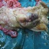
Context Neuroendocrine tumors of the pancreatic ampulla are uncommon. The final diagnosis is based on histology, and at times, it may be difficult to diagnose them pre-operatively since they present with a similar clinical picture to adenocarcinomas of this region. Objective To identify neuroendocrine tumors of the ampulla, as well as their presentation and management. Design A retrospective review of patients treated at a tertiary care institute was performed over a six-year period from 2005 to 2010. Patients Cases with periampullary cancers were investigated. Main outcome measures The case records were scrutinised for the clinical presentation, management and outcomes. Results A total of 4 cases (7.7%) of neuroendocrine tumors of the ampulla were identified from 52 patients with periampullary lesions, at a mean age of presentation of 49 years. The common mode of presentation was progressive jaundice (3 of 4 patients); pancreaticoduodenectomy was performed in 3 patients. One patient underwent palliative endoscopic stenting for metastatic disease. On histopathology, 2 of the patients had poorly differentiated (neuro)endocrine carcinoma (high grade), and 2 had well differentiated (neuro)endocrine carcinoma (1 low grade and 1 intermediate). All the tumors stained positively with chromogranin A. The patients who underwent pancreaticoduodenectomy are on regular follow-up and remain free of disease. Conclusions Neuroendocrine tumors of the ampulla are distinct entities presenting clinically with jaundice. They stain positive with chromogranin A on histopathology. Pancreaticoduodenectomy should be performed as it is associated with good outcome.Downloads
References
Selvakumar E, Rajendran S, Balachandar TG,Kannan DG,Jeswanth S, Ravicahndran S. Neuroendocrine carcinoma of the ampulla of Vater :a clinicopathogic evaluation. Hepatobiliary Pancreat Dis Int 2008;7:422-425.PIMD :18693180
Mavroudis N, Rafailadis S, Syemeonidis N, Aimoniotou E, Antonopoulos E, Evgenidis N etal Carcinoid of Ampulla of vater – a report of two cases.Act Chir Belg 2005;105:213-216.
Albores- Saavedra J, Hart A, Chable- Montero F, Henson DE. Carcinoid and high grade Neuroendocrine Carcinoma of the Ampulla of Vater- A camparitive analysis of 139 caese from Surveillance , Epidemiology , and End Results Program- A population based Study.Arch Pathol Lab Med 2010;134:1692-1696.PIMD:21043824
Jaoude WA,Lau C, Sugiyama G, Duncan A. Management of Ampullary Carcinoid tumor with Pancreaticoduodenectomy.JSCR 2010;8:4.
Beasley MB, Thunnissen FB, Hasleton P .Carcinoid tumor. In Travis WD, Brambilla E, Muller –Hermelink KH, Harris CC. Pathology and Genetics of Tumors of the lung , Thymus , Heart . Lyon, France :IARC Press; 2004:59-62.World Health Organisation Classification of Tumors ; vol 10.
Klimstra D, Modlin IR, Coppola D, Loyd R, Suster S. The pathologic classification of neuroendocrine tumors, A review of nomenclature, grading and staging systems. Pancreas 2010;39:707-12.
BosmanF,Carneiro F,Hruban R,Theise N, eds.WHO Classificationof Tumors of The Digestive System.Lyon, France:IARC Press;2010.
RindiG, Kloppel G,Couvelard A.TNM staging of midgut and hindgut (neuro)endocrine tumors :a consensus proposal including a grading system.Virchow Arch.2007;451:757-62.
Hartel M, Wente MN, Sido Bernd, Friess H, Buchler MW. Carcinoid of the ampulla of vater. Journal of Gastroenterology and Hepatology 2005;20:676-681.
Carter JT, Grenert JP, Rubenstein L, Stewart L, Way LW. Neuroendocine tumors of the Ampulla of vater. Biological behaviour and surgical management. Arch Surg 2009;144:527-531.PIMD:19528385
Klein A, Clemens J, Cameron J. Periampullary neoplasms in von Recklinghausen disease. Surgery 1989;106:815-9.
Relles D, Back J, Witkiewicz A, Yeo CJ. Periampullary and Duodenal neoplasms in neurofibromatosis type I :two cases an updated 20-year review of the literature yielding 76 cases. J Gastrointest Surg 2010;14:1052-61.PIMD:20300877
Gilani N, Ramirez FC. Endoscopic resection of an ampullary carcinoid presenting with upper gastrointestinal bleeding : A case report and review of literature.World J Gastroenterol 2007;13:168-70.PIMD:17451212
Senda E, Fujimoto K, Ohnishi K, Higashida A, Ashida C, Okutani T etal. Minute ampullary carcinoid tumor with lymph node metastasis : a case report and review of literature. World Journal Of Surgical Oncology2009;7:9. PIMD :19159493
Singhal D, Vasdev N, Soin A, Gupta S, Nundy S. Distinguishing between periampullary carcinoid and carcinomas: is this possible preoperatively.Indian J Gastroenterol 2006;25:206-7.PIMD:18693180
Karatzas G,Kouraklis G,Karayiannakis A,Patapis P,Givalos N,Kaperonis E. Ampullary and jejunal stromal tumor associated with Vion Recklinghausen’s disease presenting as gastrointestinal bleeding and jaundice. Eur J Surg Oncol 200;26:428-429.
Hatzitheoklitos E, Büchler MW, Friess H, Poch B, Ebert M, Mohr W etal. Carcinoid of the ampulla of vater , clinical characterstics and morphological features. Cancer1994 ;73:1580-1588.
Makhlouf HR, Burke AP, Sobin LH.Carcinoid of the ampulla of vater : a comparison with duodenal carcinoid tumors .Cancer 1999;85;1241-1249.
Norton JA, Kivlen M , Li M, Scheider D, Chuter T, Jensen RT. Morbidity and mortality of aggressive resection in patients with advanced neuroendocrine tumors. Arch Surg 2003;138;859- 866.
Poultides GA,Frederick WA. Carconoid of the ampulla of vater : Morphologic features and clinical implications. World J Gastroenterol.2006;12:7058-60.PIMD:17109507
Hwang S, Lee SG, Lee YJ, Han DJ, Kim SC, Kwon SH etal. Radical surgical resection for carcinoid tumors of the ampulla. J Gastrointest Surg 2008;12:713-7.
Pyun DK, Moon G, Han J, Kim MH, Lee SS, Seo DW.A carcinoid tumor of the ampulla of vater treated by endoscopic snare papillectomy. The Korean Journal of Internal Medicine 2004;19;257-260.
Sakka N, Smith RA, Whelan P, Ghaneh P, Sutton R, Raraty M etal. A preoperative score for resected pancreatic and periampullary neuroendocrine tumors.Pancreatology 2009;9;670- 676.PIMD:19684431
Jarufe NP, Coldham C, Orug T, Mayer AD, Mirza DF, Buckels JA etal. Neuroendocrine tumors of the pancreas: predictors of survival after surgical treatment. Dig Surg 2005; 22:157-162.
Hochwald SN, Zee S, Conlon KC, Colleoni R, Louie O, Brennan MF etal. Prognostic factors in pancreatic endocrine neoplasms: an analysis of 136 cases with a proposal for low-grade and intermediate grade groups. J Clin Oncol 2002; 20:2633-2642.
Copyright (c) 2014 Mayank Jayant, Rajpal Punia, Robin Kaushik, Rajeev Sharma, Atul Sachdev, Nikhil K Nadkarni, Ashok Attri

This work is licensed under a Creative Commons Attribution 4.0 International License.
As a member of Publisher International Linking Association, PILA, iMedPub Group’s JOP follows the Creative Commons Attribution License and Scholars Open Access publishing policies. Journal of the Pancreas is the Council Contributor Member of Council of Science Editors (CSE) and following the CSE slogan Education, Ethics, and Evidence for Editors.