
A Minute Pancreatic Ductal Adenocarcinoma with Lipomatous Pseudohypertrophy of the Pancreas
Abstract
Context This report describes a minute pancreatic ductal adenocarcinoma that appeared to be very early stage in the tumor progression from the study of the molecular abnormalities. Moreover, lipomatous pseudohypertrophy, a rare disease, was shown in synchrony.
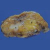
Case report A 78-year-old male was admitted to our department due to an incidental pancreatic tumor. Abdominal dynamic computed tomography showed an enlarged pancreas and diffuse fat-low density in the whole pancreas was demonstrated. In the pancreatic body, a slightly enhanced 10 mm mass in the early phase was shown. He underwent a distal pancreatectomy. The histological features of the tumor revealed abundant fibrosis and duct lesions with various atypia. Duct lesions equivalent to well differentiated adenocarcinoma were shown sparsely, but no vessel or lymphatic permeation nor perineural invasion were observed. In the background pancreas, diffuse fatty infiltrations that were composed of abundant normal adipose tissue and scattered pancreatic parenchyma were shown. The results of immunolabeling for MUC1, p16, p53, and Smad4 demonstrated that there is a possibility of coexistence of precancerous duct lesions and cancerous ones in the genetic progression of pancreatic cancer.
Conclusion Above results suggested that this pancreatic ductal adenocarcinoma with lipomatous pseudohypertrophy might be very early stage in the tumor progression.
Downloads
References
Hruban RH, Takaori K, Klimstra DS, Adsay NV, Albores-Saavedra J, Biankin AV, et al. An illustraited consensus on the classification of pancreatic intraepithelial neoplasia and intraductal papillary mucinous neoplasms. Am J Surg Pathol 2004; 28:977-87. [PMID 15252303]
Takaori K, Kobashi Y, Matsusue S, Matsui K, Yamamoto T. Clinicopathological features of pancreatic intraepithelial neoplasias and their relationship to intraductal papillary-mucinous tumors. J Hepatobiliary Pancreat Surg 2003; 10:125-36. [PMID 14505145]
Hruban RH, Klimstra DS, Pitman MB. Tumors of the Pancreas, 4th ed. In: AFIP Atlas of Tumor Pathology, Series IV. Washington (DC): Armed Forces Institute of Pathology; 2006.
Maitra A, Adsay NV, Argani P, Iacobuzio-Donahue C, De Marzo A, Cameron JL, et al. Multicomponent analysis of the pancreatic adenocarcinoma progression model using a pancreatic intraepithelial neoplasia tissue microarray. Mod Pathol 2003; 16:902-12. [PMID 13679454]
Wilentz RE, Iacobuzio-Donahue CA, Argani P, McCarthy DM, Parsons JL, Yeo CJ, et al. Loss of expression of Dpc4 in pancreatic intraepithelial neoplasia: evidence that DPC4 inactivation occurs late in neoplastic progression. Cancer Res 2000; 60:2002-6. [PMID 10766191]
Wilentz RE, Su GH, Dai JL, Sparks AB, Argani P, Sohn TA, et al. Immunohistochemistry labeling for Dpc4 mirrors genetic status in pancreatic and peripancreatic adenocarcinomas: a new marker of DPC4 inactivation. Am J Pathol 2000; 156:37-43. [PMID 10623651]
Wilentz RE, Geradts J, Maynard R, Offerhaus GJ, Kang M, Goggins M, et al. Inactivation of the p16 (INK4A) tumor-suppressor gene in pancreatic duct lesions: loss of intranuclear expression. Cancer Res 1998; 58:4740-4. [PMID 9788631]
Hruban RH, Adsay NV, Albores-Saavedra J, Anver MR, Biankin AV, Boivin GP, et al. Pathology of genetically engineered mouse models of pancreatic exocrine cancer: consensus report and recommendations. Cancer Res 2006; 66:95-106 [PMID 16397221]
Mahadevan D, Von Hoff DD. Tumor-stroma interactions in pancreatic ductal adenocarcinoma. Mol Cancer Ther 2007; 6:1186-97. [PMID 17406031]
Adsay NV, Merati K, Andea A, Sarkar F, Hruban RH, Wilentz RE, et al. The dichotomy in the preinvasive neoplasia to invasive carcinoma sequence in the pancreas: differential expression of MUC1 and MUC2 supports the existence of two separate pathways of carcinogenesis. Mod Pathol 2002; 15:1087-95. [PMID 12379756]
Ueda M, Miura Y, Kunihiro O, Ishikawa T, Ichikawa Y, Endo I, et al. MUC1 overexpression is the most reliable marker of invasive carcinoma in intraductal papillary-mucinous tumor (IPMT). Hepatogastroenterology 2005; 52:398-403. [PMID 15816444]
Hantelmann W. Fettsucht und Atrophie der Bauch specicheldruse bei Jungendlichen. Virchows Arch 1931; 282:630-42.
Yasuda M, Niina Y, Uchida M, Fujimori N, Nakamura T, Oono T, et al. A case of lipomatous pseudohypertrophy of the pancreas diagnosed by typical imaging. JOP. J Pancreas (Online) 2010; 11:385-8. [PMID 20601816]
Altinel D, Basturk O, Sarmiento JM, Martin D, Jacobs MJ, Kooby DA, Adsay NV. Lipomatous pseudohypertrophy of the pancreas. A clinicopathologically distinct entity. Pancreas 2010; 39:392-7. [PMID 19904221]
Copyright (c) 2014 Sadanobu Izumi

This work is licensed under a Creative Commons Attribution 4.0 International License.
As a member of Publisher International Linking Association, PILA, iMedPub Group’s JOP follows the Creative Commons Attribution License and Scholars Open Access publishing policies. Journal of the Pancreas is the Council Contributor Member of Council of Science Editors (CSE) and following the CSE slogan Education, Ethics, and Evidence for Editors.