
Pancreas Cystic Lymphangioma Diagnosed with EUS-FNA
Abstract
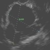
Context Endoscopic ultrasound has proved to be an invaluable tool when obtaining high quality images of the pancreas. Furthermore, fine-needle aspiration of suspected lesions can be carried out simultaneously thus providing tissue samples for cytologic diagnosis. We present two cases of a rare pancreatic lesion that were diagnosed by endoscopic ultrasound with fine-needle aspiration. Case #1 A 60-year-old asymptomatic gentleman was found to have an incidental pancreatic lesion on abdominal computed tomography scan during a cardiac workup. Patient had no personal or family medical history that would predispose him to pancreatic lesions. Endoscopic ultrasound was performed and patient was diagnosed with pancreatic cystic lymphangioma. Case #2 A 40-year-old asymptomatic gentleman with history of heavy alcohol use was found to have an incidental pancreatic lesion on computed tomography scan during a work up of chest pain. Computed tomography guided fine-needle aspiration was negative for malignancy but no other studies were performed on the fluid sample at that time. Patient was then referred to our institution after repeat computed tomography scan showed a stable lesion. Endoscopic ultrasound did not show evidence of pancreatitis and fine-needle aspiration was consistent with pancreatic cystic lymphangioma. Discussion The universally available and escalating use of computed tomography scans has led to an increased detection of incidental cystic pancreatic lesions. Pancreatic cystic lymphangiomas are a rare lesion and account for less than one percent of all pancreatic cystic lesions. These lesions are easily and accurately diagnosed by the use of endoscopic ultrasound guided fine-needle aspiration.Downloads
References
R. Sedlack, A. Affi, E. Vazquez-Sequeiros, I.D. Norton, J.E. Clain and M.J. Wiersema, Utility of EUS in the evaluation of cystic pancreatic lesions, Gastrointest Endosc 56 (2002), pp. 543–547. PMID: 12297771
L.V. Hernandez, G. Mishra, C. Forsmark, P.V. Draganov, J.M. Petersen and S.N. Hochwald et al., Role of endoscopic ultrasound (EUS) and EUS-guided fine needle aspiration in the diagnosis and treatment of cystic lesions of the pancreas, Pancreas 25 (2002), pp. 222–228. PMID 12370531
H. Abe, K. Kubota, T. Noie, Y. Bandai and M. Makuuchi, Cystic lymphangioma of the pancreas: a case report with special reference to embryological development, Am J Gastroenterol 92 (1997), pp. 1566–1567. PMID 9317093
Jathal A, Arsenescu R, Crowe G, Movva R, Shamoun DK. Diagnosis of pancreatic
cystic lymphangioma with EUS-guided FNA: report of a case. Gastrointest Endosc. 2005 Jun; 61(7):920-922. PMID 15933705
E. Paal, L.D. Thompson and C.S. Heffess, A clinicopathologic and immunohistochemical study of ten pancreatic lymphangiomas and a review of the literature, Cancer 82 (1998), pp. 2150–2158. PMID 9610694
B.C. Bounds and W.R. Brugge, EUS diagnosis of cystic lesions of the pancreas, Int J Gastrointest Cancer 30 (2001), pp. 27–31. PMID 12489578
Raddaoui E., Clinical utility and diagnostic accuracy of endoscopic ultrasound-guided fine needle aspiration of pancreatic lesions: Saudi Arabian experience. Acta Cytol. 2011; 55(1): 26-29. PMID 21135518
Sanaka M.R., Kowalski T.E. Cystic lymphangioma of the pancreas. Clinical Gastroenterology and Hepatology 2007; 5(3): 10–11. PMID 17368224
Copyright (c) 2014 Adam W Coe, John Evans, Jason Conway

This work is licensed under a Creative Commons Attribution 4.0 International License.
As a member of Publisher International Linking Association, PILA, iMedPub Group’s JOP follows the Creative Commons Attribution License and Scholars Open Access publishing policies. Journal of the Pancreas is the Council Contributor Member of Council of Science Editors (CSE) and following the CSE slogan Education, Ethics, and Evidence for Editors.