
Pancreatoblastoma: A Rare Tumor Still Evolving in Clinical Presentation and Histology
Abstract
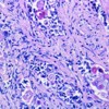
Context Pancreatoblastoma is a rare neoplasm in adults with a total of only 24 cases that have been reported in the literature. Adult pancreatoblastomas are large tumors and majority are larger than 8 cm at the time of diagnosis. Metastasis is seen in 26% of adults and usually involves the liver and then the lymph nodes. Metastasis is usually observed in cases where the primary tumor measures more than 10 cm. Pancreatoblastoma is named after its resemblance to fetal pancreatic tissue in the seventh week of life. The presence of squamoid corpuscles with a morular appearance is the most characteristic feature of the tumor. Pancreatoblastomas can have mixed features of both endocrine and exocrine cells; however, acinar differentiation is the most prevalent feature. Case report We present a case of a 27-year-old female with a 3.6 cm pancreatoblastoma with metastasis to the liver and lungs as well as to the breast. This case has several distinguishing features from previously reported cases. Such widespread metastasis is unusual given the small size of the primary tumor. Also, metastasis to the breast from a pancreatoblastoma has been previously undescribed in literature. The histological features in our case of pancreatoblastoma were atypical, characterized by the absence of acinar component, supported by the lack of staining for both trypsin and lipase in the tumor, which has not been described in literature. Additionally, the nests of squamous cells in this tumor had a pilomatricoma like morphology as opposed to the morular appearance of the squamoid corpuscles seen in classical cases. Conclusion Pancreatoblastoma can have an atypical clinical picture and a small primary with extensive metastasis to unusual sites may present a diagnostic challenge. Given its rarity, a high index of suspicion is required to correctly diagnose this condition. The histology reported on this case is unique and has not been reported in the literature.
Downloads
References
Palosaari D, Clayton F, Seaman J. Pancreatoblastoma in an adult. Arch Pathol Lab Med 1986; 110:650-2 (PMID: 3013120)
Cavallin A, Falconi M, Bortesi L, Crippa S, Barugola G, Butorini G. Pancreatoblastoma in Adults: A review of the literature. Pancreatology 2009;9:73-80 (PMID: 19077457)
Klimstra DS, wenig BM, Adair CF , Heffess CS. Pancreatoblastoma: a clinicopathologic study and review of the literature. Am J Surg Pathol 1995;19:1371-1389. (PMID: 7503360)
Charlton-Ouw KM, Kaiser CL, Tong GX, Allendorf JD, Chabot JA. J Pancreas 2008;9(6):733-738 (PMID: 18981556)
Montemarano H, Lonergan GJ, Bulas DI, Selby DM. Pancreatoblastoma: imaging findings in 10 patients and review of the literature. Radiology 2000;214:476-482 (PMID: 10671596)
Buchino JJ, Castello FM, Nagaraj HS: Pancreatoblastoma: A histochemical and ultrastructural analysis. Cancer 1984;53:963-969. (PMID: 6141001)
Rosai Juan. Rosai and Ackerman's Surgical Pathology. Ninth Edition. Mosby. Edinburgh, New York, 2004.
Sack GH Jr, Levin J, Bell, WR. Trousseau's syndrome and other manifestations of chronic disseminated coagulopathy in patients with neoplasms: Clinical, pathophysiologic, and therapeutic features. Medicine (Baltimore) 1977; 56:1.
Shaib W, Deng Y, Zilterman D, Lundberg B, Saif MW. Assessing risk and mortality of venous thromboembolism in pancreatic cancer patients. Anticancer Res. 2010 Oct;30(10):4261-4. (PMID: 21036750)
Caine GJ, Stonelake PS, Lip GY, Kehoe ST. The hypercoagulable state of malignancy: pathogenesis and current debate. Neoplasia. 2002 Nov-Dec;4(6):465-73. (PMID: 12407439)
Copyright (c) 2014 Chitra Balasundaram, Munish Luthra, Disaya Chavalitdhamrong, Jonathan Chow, Hina Khan, Paul JH Endres

This work is licensed under a Creative Commons Attribution 4.0 International License.
As a member of Publisher International Linking Association, PILA, iMedPub Group’s JOP follows the Creative Commons Attribution License and Scholars Open Access publishing policies. Journal of the Pancreas is the Council Contributor Member of Council of Science Editors (CSE) and following the CSE slogan Education, Ethics, and Evidence for Editors.