
Intraductal Oncocytic Papillary Neoplasm of the Pancreas: Report of a Case Requiring Completion Pancreatectomy
Abstract
Context Cystic tumors of the pancreas have been diagnosed with increasing frequency. Intraductal oncocytic papillary neoplasm is a rare type of cystic pancreatic tumor. Intraductal oncocytic papillary neoplasm is considered a distinct entity with the potential of developing into invasive carcinoma and it should be differentiated from other cystic tumors of the pancreas, including mucinous cystic neoplasm and other forms of intraductal papillary mucinous neoplasm (IPMN). Histologically, the formation of oncocytic cells and the complex morphology of the papillae distinguish intraductal oncocytic papillary neoplasm from IPMN. While the number of publications addressing the diagnosis, management and follow-up of patients with IPMN has been increasing, the behavior differences between IPMN and intraductal oncocytic papillary neoplasm have not been elucidated, secondary to very limited clinical experience. Case report Here, we are presenting a case of a patient with the diagnosis of intraductal oncocytic papillary neoplasm of the pancreas developing into invasive cancer. Conclusion This case stresses the necessity for lifelong surveillance of the remnant pancreas following partial pancreatectomy for intraductal oncocytic papillary neoplasm, due to the risk of developing multifocal disease.
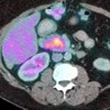
Image: PET CT image shows diffuse uptake of contrast in the head of the pancreas.
Downloads
References
Adsay NV, Adair CF, Heffess CS, Klimstra DS. Intraductal oncocytic papillary neoplasms of the pancreas. Am J Surg Pathol 1996; 20:980-94.
Chari ST,YadavD, Smyrk TC, et al. Study of recurrence after surgical resection of intraductal papillary mucinous neoplasm of the pancreas. Gastroenterology. 2002;123:1500–1507.
Matthaei H, Norris AL, Tsiatis AC, et al. Clinicopathological characteristics and molecular analyses of multifocal intraductal papillary mucinous neoplasms of the pancreas. Ann Surg. 2012;255:326-33.
Xiao HD, Yamaguchi H, Dias-Santagata D, et al. Molecular characteristics and biological behaviours of the oncocytic and pancreatobiliary subtypes of intraductal papillary mucinous neoplasms. J Pathol. 2011;224:508-16.
Fischer MA, Donati O, Heinrich et al. Intraductal oncocytic papillary neoplasm of the pancreas: a radio-pathological case study. JOP 2010;11:49-54.
Liszka L, Pajak J, Zielinska E et al. Intraductal oncocytic papillary neoplasms of the pancreas and bile ducts: a description of five new cases and review based on a systemic survey of the literature. J Hepatobiliary Pancreat Sci 2010;17:246-261.
D'Angelica M, Brennan MF, Suriawinata AA, et al. Intraductal papillary mucinous neoplasms of the pancreas: an analysis of clinicopathologic features and outcome. Ann Surg. 2004;239:400-8.
Sohn TA, Yeo CJ, Cameron JL, et al. Intraductal papillary mucinous neoplasms of the pancreas: an updated experience. Ann Surg. 2004;239:788-97; discussion 797-9.
Murakami Y, Uemura K, Sudo T, et al. Invasive intraductal papillary-mucinous neoplasm of the pancreas: comparison with pancreatic ductal adenocarcinoma. J Surg Oncol. 2009;100:13-8.
Ishida M, Egawa S, Aoki T, et al. Characteristic clinicopathological features of the types of intraductal papillary-mucinous neoplasms of the pancreas. Pancreas. 2007;35:348-52.
Zen Y, Sasaki M, Fujii T, et al. Different expression patterns of mucin core proteins and cytokeratins during intrahepatic cholangiocarcinogenesis from biliary intraepithelial neoplasia and intraductal papillary neoplasm of the bile duct—an immunohistochemical study of 110 cases of hepatolithiasis. J Hepatol. 2006;44:350-8.
Copyright (c) 2014 Max V Wohlauer, Martine McManus, Norio Fukami, Csaba Gajdos

This work is licensed under a Creative Commons Attribution 4.0 International License.
As a member of Publisher International Linking Association, PILA, iMedPub Group’s JOP follows the Creative Commons Attribution License and Scholars Open Access publishing policies. Journal of the Pancreas is the Council Contributor Member of Council of Science Editors (CSE) and following the CSE slogan Education, Ethics, and Evidence for Editors.